
Hilo Medical Center
Imaging Department
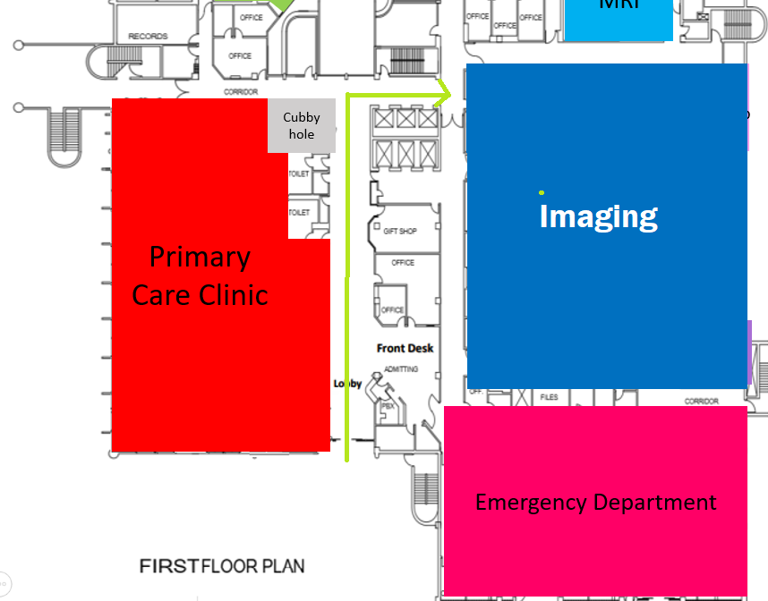
Hilo Medical Center’s Imaging Department is responsible for providing and managing the hospital’s inpatient and outpatient imaging services. The department provides patients with the best possible care, using state-of-the-art equipment in a professional, caring and timely manner. In 2015, the Imaging Department underwent renovation and upgrade.
Services offered consists of 6 modalities:
Angiography is the imaging of blood vessels using contrast injected into the blood stream to determine the presence of blockages or clots in arteries or veins and to diagnose patient heart functions. In 2015, Hilo Medical Center constructed a brand new angiography suite to house the new angiography equipment, a Siemens Artis Zee Biplane system that features excellent performance for imaging and position flexibility. This equipment is ideal for a variety of uses for cardiology, interventional radiology and vascular surgery.

CT is an advanced form of X-ray technology that creates multiple X-rays per scan. Hilo Medical Center’s uses a 32-slice CT machine, which involves a patient lying on the CT table, slowly passing through the center of a large, doughnut-shaped X-ray machine. Some tests require contrast dye. In 2015, we installed a new Toshiba Aquilion One Vision 640-slice CT scanner. Thanks to state Trauma Program funds, our Imaging Department will be equipped with the best CT scanner on the market. In just 0.275 second and one revolution, we’ll be able to scan the entire heart. The high-quality images and scanning speed mean more accurate diagnosis for patients, especially for children or those who are in a great deal of pain.

Diagnostic X-ray/Fluoroscopy uses minimal amounts radiation. X-ray beams are absorbed in different amounts depending on the density of the tissue they pass through the body. The leftover x-rays that pass through your body are captured X-ray film. Some tests require contrast dye.

MRI uses a large magnet and radio waves to examine organs and tissues. Patients, lying on a table, slowly enter the large, tube-shaped MRI machine. Patients are offered headphones with soothing music to drown out the constant thumping sounds. The magnets and radio waves create cross-sectional MRI images. The MRI machine can also produce 3-D images that may be viewed from different angles.

Nuclear Medicine uses safe, radioactive materials, known as tracers, to create precise images of organs function, analyze biological specimens and treat disease. Tracers may be ingested, inhaled, or injected. The amount of radiation received from nuclear imaging is around the same amount as an X-ray.
Ultrasound is an imaging method that uses high-frequency sound waves to view the heart, blood vessels, kidneys, liver and other organs.
During pregnancy, doctors use ultrasound tests to examine the fetus. Unlike x-rays, ultrasound does not involve exposure to radiation.
